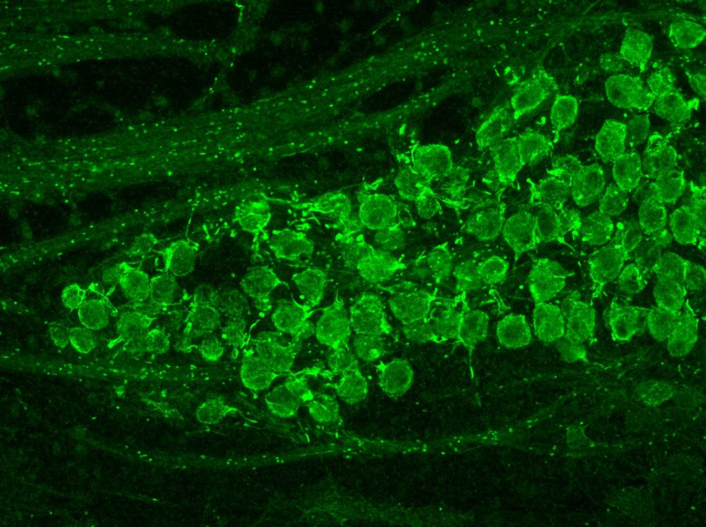
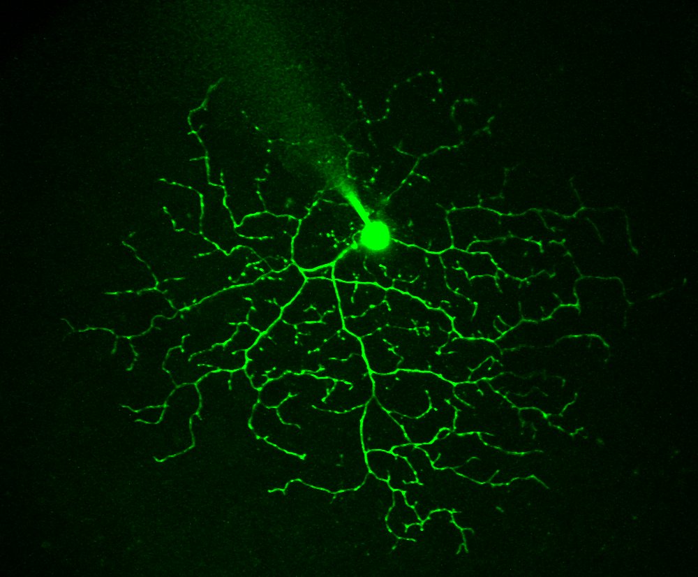
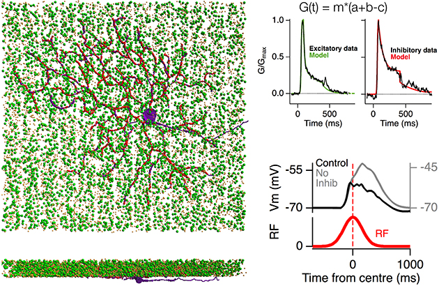
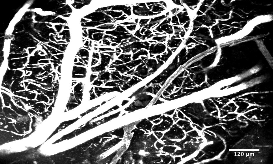
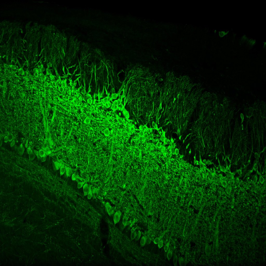
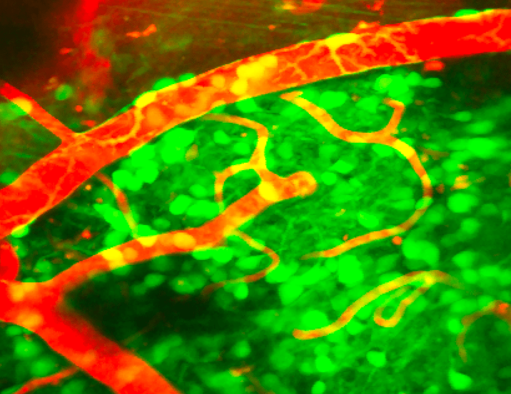
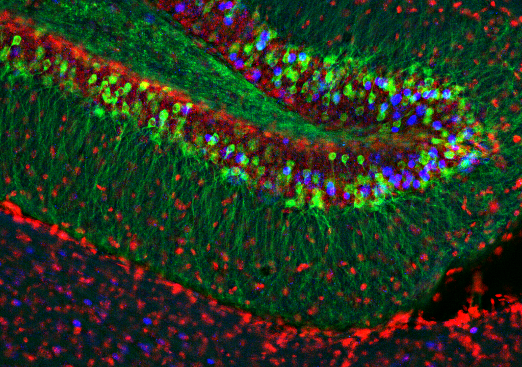
Kv3.1 in the MNTB, note the Kv1.3 puncta in the node of ranvierAmygdalar neurons responding to stimulation of cortical inputsFirst image taken on our custom built muti-photon microscope (Cardiac myocyte)A retinal ganglion cell filled with Alex-488 through the patch pipetteCircuit model of the retina exploring motion anticipationBrain vasculature in the olfactory bulb, labelled with TexasRed-DextranFreeze-fracture electron micrograph of the glomerular layer of the olfactory bulbPresynaptic patch-clamp recording from the Calyx of held in an acute brain slicePG cells responding to respiration and odour stimulationDifferent type of Periglomerular cells: Tyrosine Hydroxylase (Cyan), Calbindin (Yellow) and Calretinin (Magenta)The inside of the 1st Ti:Sapphire laser I used (Mira 900)Mitral and Tufted cell expressing GCaMP6The synaptic terminals of retinal bipolar cells responding to moving stimuliMy BrainReceptve field of a retinal ganglion cell mapped on a multi-electrode array with FBPNestin labelled PG cells and pericytes in green with the vasculature in red.Dentate Gyrus