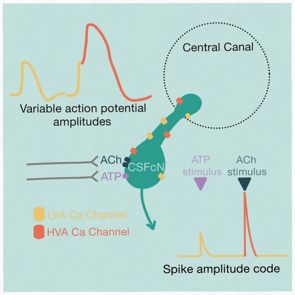
This project explored the physiological properties of the intriguing cerebrospinal fluid contacting neurons within the spinal cord. These sensory neurons are able to sense bending of the spinal cord and are involved in regulating motor output from the spinal cord. We showed that these sensory neurons have unusual action potentials, rather than using the typical sodium channels for depolarisation they use calcium channels.

We showed that CSFcNS use 2 different types of Ca channels that enables them to fire spikes of different amplitude, they can use this amplitude code to signal inputs from different neurotransmitters. This is dramatically different from how most other neurons operate, where action potentials are all-or-non and generate a binary code. However, the graded action potentials of cerebrospinal fluid contacting neurons do bear similarities to other sensory neurons found in the retina. The full paper can be viewed here.
CSFcNs release GABA from their endbulb
Some follow-up work that didn’t make it to the paper…
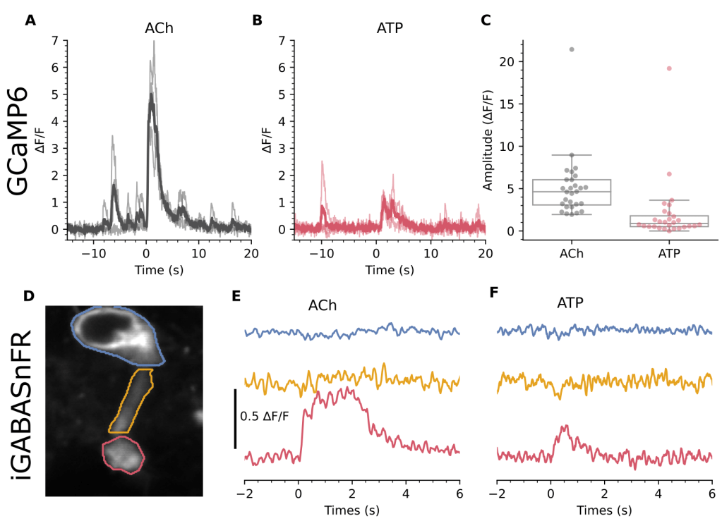
In our paper, we show that ACh and ATP evoked different amplitude Ca2+ responses in CSFcNs (Fig. 7 A-C, reproduced below). In subsequent experiments we used iGABASnFR expressed in CSFcNS (panel D) and are possibly the first to show that CSFcNs release GABA from their endbulb upon stimulation with ACh (panel E) or ATP (panel F).
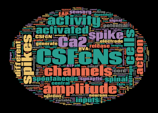
Intriguing cells!
