And its application to multi-neuronal electrophysiology and imaging
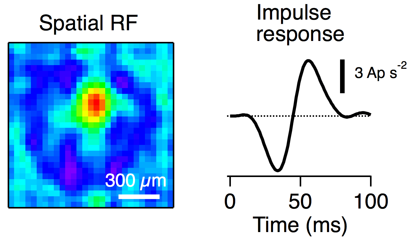
Neurons in sensory systems have receptive fields. In the somatosensory system, this might be an area of the skin to which the neuron is responsive, whereas in the visual system the receptive field is an area on the retina. Several properties of the receptive field are of interest. The spatial extent of the receptive field, e.g. the area on the retina to which the neuron is responsive, and the temporal characteristics, e.g. when will the neuron respond after applying a stimulus. Measuring the properties of a neuron’s receptive field is an important step towards understanding its function.
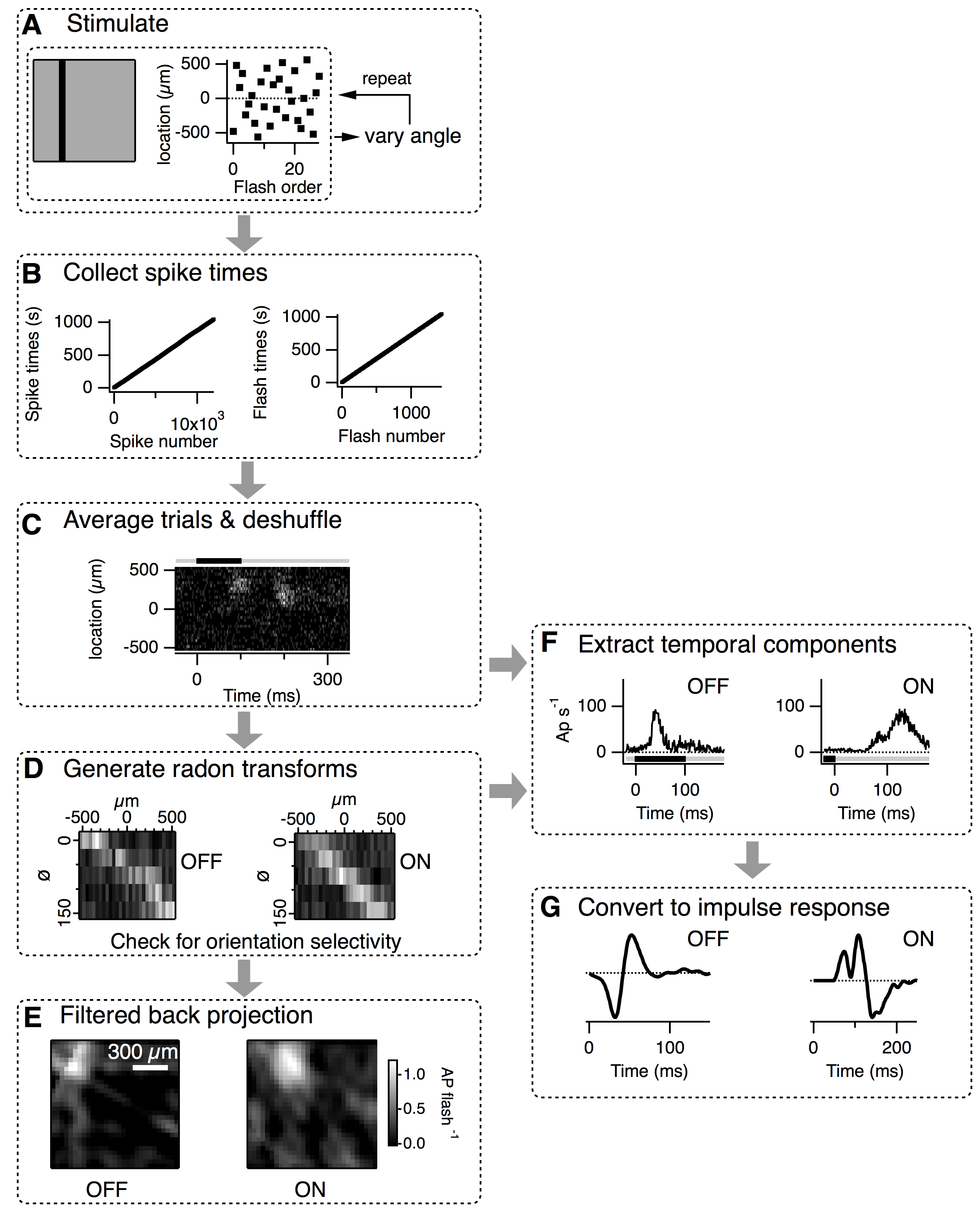
This project developed a new method that allows the rapid mapping of receptive fields from multiple neurons simultaneously. The procedure is illustrated to the right and the full details are available in the paper here. A series of bars are flashed at different locations across the retina and this is repeated with the bars rotated to cover at least 5 evenly spaced angles. The receptive fields can then be easily recovered using an algorithm applied in CAT scans, the filtered back projection. The code to implement this method is available here.
We showed that the FBP method can recover both the spatial and temporal components of the receptive field. The FBP method had several advantages over the frequently used “spike-triggered average” approach. The FBP could recover receptive fields significantly faster, with higher signal to noise and the resolution of the temporal impulse response was superior.
We also demonstrated how this method is suitable for functional imaging of neural activity. Imaging data is often noisy and clear unitary events such as spikes are not readily discernible. As our method does not rely on detection of events it readily lends itself to mapping receptive fields of neurons measured in imaging experiments. We demonstrated this by mapping the receptive fields of an array of retinal bipolar cell synapses expressing SyGCaMP6 shown below.
